

Medically reviewed by
USA Fibroid Centers Medical Review Team
Experienced interventional radiology specialists
Reviewed: July 2026
Adenomyosis does not have a universally accepted numbered staging system, the way endometriosis does. While you may have heard terms like “Stage 1” or “Stage 4” used in online discussions, no global medical standard formally assigns those labels to adenomyosis. Instead, specialists assess the condition using a different framework, focusing on where the tissue is located and how deeply it has grown into the uterine muscle. Knowing this distinction may help you understand your diagnosis and the next steps your doctor recommends.
What Adenomyosis Is (and Why It’s Hard to Stage)
Adenomyosis occurs when the inner lining of the uterus, called the endometrium, grows into the muscular wall of the uterus, known as the myometrium. This displaced tissue continues to thicken, break down, and bleed during each menstrual cycle, which can cause the uterus to enlarge and become inflamed.
Unlike cancer, which specialists assess based on how far it has spread to other organs and tissues, adenomyosis remains confined within the uterus. This makes a traditional staging framework less clinically useful. Unlike endometriosis, which can spread to the ovaries, fallopian tubes, and other structures outside the uterus, adenomyosis does not travel beyond the uterine wall.
Since the disease stays within one organ and varies so much from person to person, the medical community has not reached a consensus on a single classification system. Research published in the Journal of Clinical Medicine confirmed that as of 2023, no universally validated staging system for adenomyosis has been compiled.[1] Several systems have been proposed, but none have been adopted globally.
How Specialists Actually Classify Adenomyosis
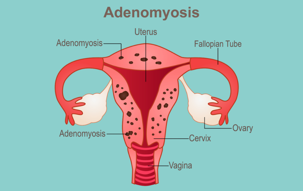
Rather than assigning a stage number, specialists evaluate two key factors: where the endometrial tissue is located and how deeply it has invaded the uterine muscle. These two dimensions help characterize the condition and guide treatment decisions.
Focal Adenomyosis
Focal adenomyosis refers to endometrial tissue concentrated in one area of the uterine wall. It may appear as a localized mass, sometimes called an adenomyoma. Focal adenomyosis can cause significant symptoms such as localized pelvic pain, heavy or prolonged menstrual bleeding, chronic pelvic pressure, and painful intercourse. Still, because it is contained to a specific region, imaging tends to identify it more precisely.
Diffuse Adenomyosis
Diffuse adenomyosis involves endometrial tissue spread throughout a larger portion of the uterine wall. This form is associated with a more uniformly enlarged uterus and may be linked to more consistent, widespread symptoms. Women with diffuse adenomyosis often experience heavy menstrual bleeding and pelvic pain more frequently.
What Imaging Reveals
Transvaginal ultrasound (TVUS) is typically the first imaging tool used when adenomyosis is suspected. It is widely available, non-invasive, and can identify signs such as an asymmetrically thickened uterus, myometrial cysts, and irregular tissue patterns.
Magnetic resonance imaging (MRI) is used as a second-line tool when ultrasound findings are unclear or when a more detailed view of the myometrium is needed. An MRI can measure junctional zone thickness, with a measurement of 12 mm or more considered a key diagnostic marker.[2] These imaging findings help specialists understand the extent and pattern of the condition without relying on a numbered stage.
Does Adenomyosis Progress Over Time?
Adenomyosis is an estrogen-dependent condition. It tends to develop during the reproductive years and, for many women, symptoms ease after menopause when estrogen levels decline. However, the course of the condition varies widely between individuals.
Some women experience mild, manageable symptoms that do not intensify significantly. Others find that symptoms become more disruptive over time, particularly if the condition is left unaddressed. Factors such as the extent of tissue involvement, overall hormonal activity, and the presence of related conditions like uterine fibroids can all influence how symptoms evolve.
The important takeaway is that progression is not guaranteed, nor is it the same for everyone. Symptoms that begin as occasional pelvic pressure or slightly heavier periods may stay at that level for years, or they may shift. Consulting with an interventional radiologist or specialist when symptoms change can help you stay ahead of the condition rather than simply responding to it.
Living with adenomyosis can be painful. Understanding the causes of adenomyosis pain can help recognize when it may be time to seek professional evaluation and treatment.
Symptoms That May Signal a More Significant Condition
Since adenomyosis does not use a numbered staging framework, symptoms play an important role in helping both patients and specialists understand the condition’s current impact. Symptoms alone cannot confirm the degree of tissue involvement, but they do help paint a fuller picture alongside imaging.
Common adenomyosis symptoms include:
- Heavy or prolonged menstrual bleeding
- Severe menstrual cramps that interfere with daily activities
- Persistent or intermittent pelvic pain outside of the menstrual cycle
- Painful intercourse
- A feeling of pelvic pressure or fullness
- An enlarged, tender uterus detected during a pelvic exam
Symptoms that significantly affect your quality of life, work, or daily routine are a signal worth taking seriously. An accurate diagnosis through imaging is the only way to understand the specific pattern and extent of the condition in your uterus.
It is also worth noting that adenomyosis frequently coexists with other uterine conditions. If you have been told you have fibroids and are still experiencing significant symptoms, it is worth asking your specialist whether adenomyosis and fibroids may both be contributing.
Getting an Accurate Diagnosis
Adenomyosis symptoms overlap with other uterine conditions, which is why diagnosis requires more than a symptom review. A pelvic exam may reveal an enlarged or tender uterus, which can prompt further investigation. From there, transvaginal ultrasound is typically the first imaging step.
If TVUS findings are inconclusive, your specialist may recommend an MRI for a more detailed view of the myometrial wall and junctional zone. These imaging tools allow specialists to assess whether adenomyosis is focal or diffuse, how deep the tissue has grown into the uterine muscle, and whether any co-occurring conditions are present.
Historically, adenomyosis could only be confirmed through pathological examination following a hysterectomy. Advances in imaging technology have made it possible to identify the condition non-invasively, allowing more women to receive an accurate diagnosis while their uterus is still intact and treatment options remain open. If you have overlapping symptoms, reviewing adenomyosis vs. endometriosis may help clarify what your specialist is evaluating.
What Non-Surgical Options Are Available?
For women who want to address adenomyosis without removing the uterus, uterine artery embolization (UAE), sometimes referred to as uterine fibroid embolization (UFE) when used for fibroid-related symptoms, is a minimally invasive option that works by reducing blood flow to the affected uterine tissue. This can help shrink the displaced tissue and reduce symptoms. The procedure is performed on an outpatient basis, uses imaging guidance, and does not require general anesthesia.
Understanding how adenomyosis is classified, whether focal or diffuse, is one factor that specialists consider when evaluating whether UAE may be appropriate for a patient. An evaluation by an interventional radiologist can help determine whether this approach fits your specific case. For a closer look at this option, see our article on UFE for adenomyosis.
Treatment option: UAE/UFE
Understanding Your Condition Is the First Step. The Next Is Talking to a Specialist.
Knowing that adenomyosis does not follow a numbered staging system can feel both clarifying and unsettling. The condition is real, your symptoms are valid, and imaging-based evaluation is how specialists determine the most appropriate next steps for your individual case.
Interventional radiologists at USA Fibroid Centers evaluate each case individually using advanced imaging. For women seeking options that do not require removing the uterus, UAE is a minimally invasive, outpatient procedure with no general anesthesia and a short recovery time.
Frequently Asked Questions About Adenomyosis Stages
- Bourgioti, C., et al. “Adenomyosis: An Updated Review on Diagnosis and Classification.” Journal of Clinical Medicine 12, no. 14 (2023): 4828. mdpi.com/2077-0383/12/14/4828
- Bergeron, C., et al. “MRI for Adenomyosis: A Pictorial Review.” Insights into Imaging (2017). PMC5707223. pmc.ncbi.nlm.nih.gov/articles/PMC5707223/
- Dason, E.S., et al. “Guideline No. 437: Diagnosis and Management of Adenomyosis.” Journal of Obstetrics and Gynaecology Canada (2023). doi:10.1016/j.jogc.2023.04.008
- Benetti-Pinto, C.L., et al. “Recent Advances in Understanding and Managing Adenomyosis.” F1000Research (2019). PMC6419978. pmc.ncbi.nlm.nih.gov/articles/PMC6419978/
Medical Disclaimer: This article provides educational information about adenomyosis and available treatment options. It is not a diagnosis or medical advice. Only a qualified fibroid specialist can determine whether you have adenomyosis or recommend appropriate treatment. If you have heavy periods, pelvic pain, or other symptoms, consult a healthcare provider or contact USA Fibroid Centers for an evaluation.