

Medically reviewed by
Dr. Jacob White, MD, Interventional Radiologist, Fibroid Specialist and Medical Director
Fibroid diagnosis and non-surgical treatment
Reviewed: September 2026
A calcified fibroid is a fibroid that has hardened after losing its blood supply, and it will not go away on its own once this happens. Many women learn about fibroid calcification only after an ultrasound or MRI turns up the word “calcified” on a report, and the term alone can sound more alarming than it is. Here is what this stage of a fibroid’s life cycle actually means for your body and your treatment options.
What Is Fibroid Calcification?
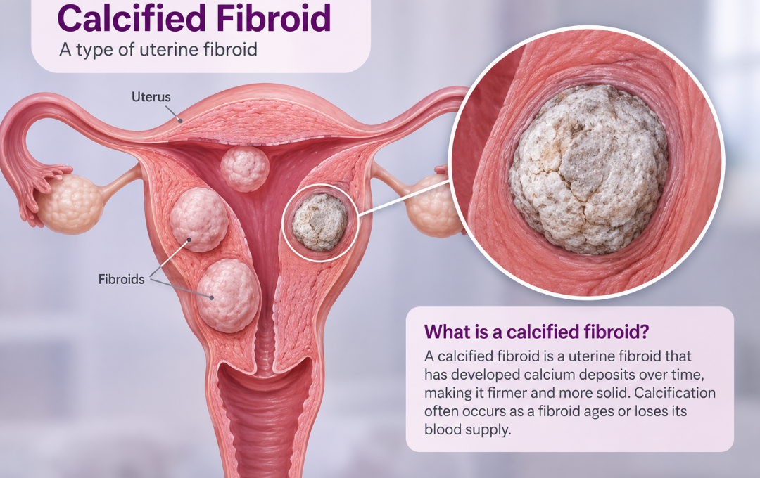
Fibroid calcification is a hardening process that some uterine fibroids go through over time. Instead of staying soft, the fibroid tissue develops calcium deposits and becomes firm, sometimes described as bone-like or rock-like. This happens in an estimated 3 to 10 percent of fibroid cases.[1]
Calcification can affect just the outer edge of a fibroid or, less often, spread through the entire growth. It is considered a form of fibroid degeneration, meaning the fibroid has reached a point where it is no longer actively growing.
Why Do Fibroids Calcify?
Fibroids depend on a steady blood supply to keep growing. When that supply is reduced or cut off, the tissue at the center of the fibroid starts to break down. Calcium deposits gradually replace the breakdown tissue, hardening the fibroid over time.[2]
This can happen for a few reasons:
- Age and menopause: Declining estrogen levels can reduce blood flow to fibroids, and calcification becomes more common in women who are approaching or past menopause
- Outgrowing blood supply: A fibroid that grows quickly may outpace the blood vessels feeding it, causing the center to degenerate and calcify while other parts may stay active
- Previous fibroid treatment: Fibroids that have been treated with uterine fibroid embolization (UFE) can calcify as they shrink, which is generally viewed as an expected part of the healing process
Understanding the different types of fibroids can help explain why some calcify while others do not, since location and size both play a role.
Different Types of Fibroids
Uterine fibroids are classified by where they grow within or on the walls of the uterus. Their location determines the specific symptoms they cause, such as heavy bleeding, pelvic pressure, or pain.
Intramural Fibroids: The most common type, growing within the muscular wall of the uterus. As they expand, they can make the uterus feel larger and cause heavier periods or dull pelvic aches.
Subserosal Fibroids: These project outward onto the exterior wall of the uterus. Because they extend into the pelvic cavity, they often press against nearby organs, causing back pain, constipation, or frequent urination.
Submucosal Fibroids: These grow just beneath the inner lining of the uterine cavity. Though less common, even small submucosal fibroids can cause severe, heavy menstrual bleeding and spotting between cycles.
Pedunculated Fibroids: These attach to the inside or outside surface of the uterus by a thin stalk, called a peduncle. If a pedunculated fibroid twists on its stalk, it can cause sudden, sharp pelvic pain.
Symptoms of Calcified Fibroids
A calcified fibroid can cause the same symptoms as other uterine fibroids, though some women notice no symptoms at all. Reported symptoms include:
- Pelvic pain or pressure
- Frequent urination
- Lower back pain
- Bloating or abdominal fullness
- Constipation or bowel discomfort
- Heavy menstrual bleeding, in some cases tied to earlier fibroid activity, and possibly linked to anemia caused by fibroids
Small calcified fibroids that are not pressing on nearby organs may go unnoticed until they show up on imaging done for another reason. Larger calcified fibroids are more likely to cause pressure-related symptoms, since size and location matter more than calcification status when it comes to how a fibroid feels day to day.
Will a Calcified Fibroid Go Away on Its Own?
No, a calcified fibroid will not go away or shrink further on its own. Calcification is already a sign that the fibroid has reached the end of its growth phase, but the hardened tissue itself typically remains in place indefinitely.[1]
This does not mean every calcified fibroid needs treatment. If a calcified fibroid is small and is not causing symptoms, many fibroid specialists recommend monitoring it with periodic imaging rather than treating it right away. Watching and waiting becomes less appropriate once a calcified fibroid starts to press on the bladder, bowel, or pelvic nerves, or if bleeding and pain continue.
Can Calcified Fibroids Turn Cancerous?
Calcified fibroids are not a sign of cancer, and calcification does not raise the already low risk of a fibroid becoming cancerous. Uterine fibroids, including calcified ones, are almost always benign. The risk of a fibroid turning into a rare cancer called leiomyosarcoma is well under one percent.[3]
In fact, some fibroid specialists view calcification on imaging as a reassuring sign, since it points to a stable, non-growing mass rather than an actively changing one. Rapid new growth, not calcification, is the pattern that typically prompts closer evaluation for something other than a fibroid.
Treatment Options for Calcified Fibroids
Treatment for a calcified fibroid depends on whether it is causing symptoms, along with its size, location, and how it is affecting your daily life.[2] Options a fibroid specialist may discuss include:
Monitoring: For calcified fibroids that are not causing symptoms, regular imaging can track the fibroid over time without immediate treatment.
Symptom management: Pain relievers or other conservative measures may help manage discomfort tied to pressure symptoms while a treatment plan is being considered.
Uterine Fibroid Embolization (UFE): UFE is a minimally invasive, non-surgical procedure that blocks blood flow to fibroids that still have an active blood supply. Fibroids with mixed calcified and non-calcified tissue may still respond to UFE, since active portions of the fibroid can shrink once blood flow is reduced. Fully calcified fibroids that no longer have meaningful blood flow are less likely to respond to UFE, which is why an accurate diagnostic workup matters before deciding on next steps. This can matter most for larger fibroids, where size and blood supply both affect how well treatment works.
A fibroid specialist can review your imaging and symptoms to help determine whether UFE, monitoring, or another approach fits your situation.
Treatment option: UFE
Understanding Your Calcified Fibroid Is the First Step
A calcified fibroid marks the end of a fibroid’s growth cycle rather than the start of a new problem, and it is not linked to cancer. Still, calcified fibroids can press on nearby organs and cause real symptoms that deserve attention.
Calcification means a fibroid has stopped growing, but it does not mean your symptoms have to stay the same. A fibroid specialist can review your imaging, explain what your specific diagnosis means, and discuss whether a minimally invasive option like UFE fits your situation.
FAQs About Fibroid Calcification
- Society of Interventional Radiology, “Uterine Fibroid Embolization,” sirweb.org/patients/uterine-fibroids/
- National Institute of Environmental Health Sciences (NIEHS), “Uterine Fibroids,” niehs.nih.gov/health/topics/conditions/uterine-fibroids
- Office on Women’s Health, U.S. Department of Health and Human Services, “Uterine Fibroids,” womenshealth.gov/a-z-topics/uterine-fibroids
Medical Disclaimer: This article provides educational information about uterine fibroids and treatment options. It is not a diagnosis or medical advice. Only a qualified fibroid specialist can determine whether you have fibroids or recommend appropriate treatment. If you have heavy periods, pelvic pain, or other symptoms, consult a healthcare provider or contact USA Fibroid Centers for an evaluation.