Diagnosing Uterine Fibroids with Ultrasound
Many women wonder, “Can fibroids be missed on an ultrasound?” The truth is that fibroids, non-cancerous growths that attach to the uterine lining or outside the uterus, are often found during a routine pelvic exam. Your doctor may then order an ultrasound to confirm the diagnosis and determine the size of the fibroids.
This article will discuss ultrasounds and how accurate they are for a uterine fibroid diagnosis.
If you suspect you have fibroids, an accurate diagnosis is important because fibroids can press on other organs and cause symptoms that can impact your life, including affecting your ability to get pregnant and causing complications during pregnancy. While many women are unaware they have fibroids, others experience symptoms such as heavy bleeding or pain.
Use our fast, free fibroid symptom checker if you think you might have uterine fibroids.
How Ultrasounds Detect Fibroids
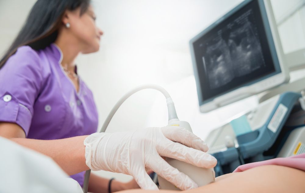
During a uterine fibroid ultrasound, the technician uses a handheld instrument called a transducer. Gel is applied to the abdomen, and the transducer is moved over the area. Sound waves travel to the uterus and create an image showing the location and size of the fibroids. The fibroids on an ultrasound appear as lighter or darker masses contrasting to the uterus.
Schedule a Consultation Online
Will Uterine Fibroids Show Up on Ultrasound?
As a safe, pain-free diagnostic tool, an ultrasound offers many advantages for detecting uterine fibroids. Ultrasound is considered the gold standard imaging test to confirm the existence of fibroids because it allows the doctor to distinguish fibroids from other conditions, such as adenomyosis, polyps, ovarian tumors, and a pregnant uterus.
However, not all fibroids may be visible on an ultrasound. For example, sound waves may have difficulty traveling through dense areas like bone or when gas is present in your intestine (colon). If this happens, the doctor may order an MRI for a fibroid diagnosis.
What Is the Best Imaging for Fibroids, Ultrasound or MRI?
An MRI, or magnetic resonance imaging, provides more detail about fibroid size and location. It can also identify different types of tumors, which can help rule out an inaccurate diagnosis. During an MRI, magnets force the protons in the body to align, after which a radiofrequency current is sent through the patient to stimulate the protons and cause them to spin. The magnetic properties of the protons tell physicians what types of tissues are present, allowing them to distinguish between abnormal tissues (such as fibroids) and healthy tissues.
A doctor may recommend an MRI for women nearing menopause or those with a larger uterus. An MRI is often used as a second option if the ultrasound does not provide enough information. The average duration of an MRI is 45 to 60 minutes. Not every woman needs an MRI; an ultrasound is usually enough to confirm a diagnosis of uterine fibroids.
Types of Ultrasound Scan to Diagnose Fibroids
Two types of ultrasound scans are useful in the diagnosis of fibroids. The first is an abdominal ultrasound scan, which moves over the abdomen to transmit sound waves. This type of ultrasound is commonly used during pregnancies to produce an image of the baby.
The second is a transvaginal ultrasound scan, which uses a small probe inserted into the vagina. This procedure is similar to a regular pelvic exam but less invasive, as the ultrasound probe is small and generally causes less discomfort than a standard exam.
What Can I Expect from a Fibroid Ultrasound?
The abdominal ultrasound is virtually painless. The transvaginal ultrasound may cause some discomfort, but it is usually less uncomfortable than a regular pelvic exam. In either case, you can return to normal activities after completing the test.
Preparing Yourself for the Procedure
Many ultrasounds require no special preparations, but there can be exceptions. Ask your doctor what you need to do to prepare for your ultrasound. Common instructions include:
- Wearing loose, comfortable clothing.
- Leaving valuables at home, as you may be asked to remove jewelry.
- Drinking water beforehand (a full bladder can provide a better view of the uterus and make it easier to see fibroids on an ultrasound).
How Long Does a Fibroid Ultrasound Procedure Take?
A fibroid ultrasound generally takes about 15 minutes. However, you will need to check in and possibly fill out some paperwork before being taken back to begin the ultrasound. After the procedure, the doctor will read the images immediately and discuss treatment.
Fibroids can have a negative impact on your life. They may affect your career, relationships, physical health, fertility, and even pregnancy. However, even if you are diagnosed with fibroids, treatment is available to help alleviate the symptoms and allow you to live a normal life.
Visit USA Fibroid Centers for Fibroid Treatment
If you are concerned about fibroids, visit our state-of-the-art outpatient clinic nearest you to speak with a fibroid specialist. We can provide an ultrasound test on-site to provide an accurate diagnosis.
If you have or are diagnosed with uterine fibroids, USA Fibroid Centers provides a safe, non-surgical treatment known as Uterine Fibroid Embolization (UFE). This minimally invasive procedure uses a tiny catheter to target embolic agents through the artery attached to the fibroid, which allows the fibroid to shrink and die.
Schedule an appointment with USA Fibroid Centers at one of our more than 40 locations nationwide to learn more about this procedure and how it can help you. Set up a consultation online with our fibroid specialists or call us at 855.615.2555 to get your questions answered.


