We have the technology and tools that allow us to see a detailed map of the body without ever touching a scalpel. Advanced technologies using diagnostic imaging, allow doctors to thread catheters throughout the body to treat a variety of conditions and diseases without invasive surgery. These images allow the doctors to have a clear picture of your fibroids to understand their location and size and offer an individual diagnosis and treatment plan.
So, how do doctors use fibroid mapping to treat conditions like uterine fibroids?
Fibroid Mapping

Understanding the below tools opens up a new world of safe, effective ways to treat these benign, uterine tumors.
At USA Fibroid Centers, our interventional radiologists (IR) use minimally invasive techniques such as ultrasounds and fluoroscopies to locate uterine fibroids. Fibroid mapping allows the doctor to diagnose and determine the number, size, location, and fibroid type. Uterine fibroids are most common in a woman’s reproductive years and present with a variety of symptoms, including heavy and abnormal bleeding, pain, pressure, infertility, and recurrent pregnancy loss.
Once located, the IR doctors can use the body’s natural pathways, the arteries, to treat the fibroids with a nonsurgical procedure called Uterine Fibroid Embolization (UFE).

Fluoroscopy
Fluoroscopy is a large device with a movable arm. This type of medical imaging shows a continuous, live X-ray on a monitor screen.
During a Uterine Fibroid Embolization procedure, the fluoroscopy passes an X-ray beam through the body focusing on the arteries leading to the uterus. The image is then transmitted to the monitor so the movement of the catheter through the arteries, as well as the contrast agent also known as “X-ray dye”, can be seen through the body.
Fluoroscopy can be used before and during the Uterine Fibroid Embolization procedure. During the scan, the patient lies on an open examination table with the fluoroscopy positioned next to the table. The arm is then moved above the body. Unlike MRIs and CAT scans, your body is not encompassed and you do not feel enclosed.
Ultrasound
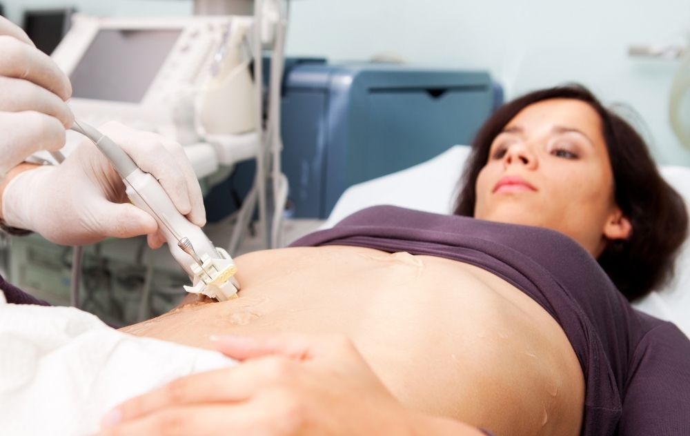
An ultrasound scan uses high-frequency sound waves to create images of the inside of the body, also known as fibroid mapping. Ultrasounds are typically used in diagnosing a pregnancy because no radiation is used. Ultrasound scans are a safe way to diagnose fibroids because they use sound waves or echoes to make an image. The sonographer applies a small amount of gel which acts as a conductive medium. Then, they use a hand-held device, also known as a transducer, which is placed on the patient’s skin. When the transducer is rubbed across the skin, an image is produced, which enables the sonographer or interventional radiologist to take pictures of the inside of the uterus.
An ultrasound’s sound waves easily travel throughout soft tissue and fluids. Ultrasounds can detect abnormal objects or tumors when the sound waves bounce off these denser surfaces. When a sound wave echos off a uterine fibroid, a darker picture is produced and you can view them on the screen. Sonographers perform ultrasound scans, which are then interpreted by an interventional radiologist which will determine the next steps and if treatment is needed.
Magnetic Resonance Imaging (MRI)
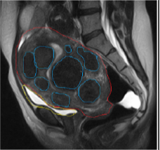
MRI uses powerful magnets and radio waves to make detailed pictures inside your body. When you have an MRI scan, you lie face-up on a table, that is slowly inserted into a large cylinder-like machine. The doctor then watches a screen inside a separate room while the MRI scan is being completed.
An MRI scan utilizes safe, radiofrequency pulses that re-align hydrogen atoms that naturally exist within the body. As the hydrogen atoms return to their usual alignment, they emit different amounts of energy depending on the type of body tissue they are in. The MRI captures this energy and creates a picture of the tissues scanned based on this information.
A computer then processes the information and generates a series of images. The images can then be studied from different angles by the interventional radiologist.
Your doctor may recommend an MRI scan if the ultrasound pictures do not show a detailed-enough picture. Most fibroids can be located with an ultrasound; however, your doctor may want an MRI picture before treatment to make a more accurate diagnosis.
UFE Treatment
Uterine Fibroid Embolization (UFE) is a minimally-invasive, FDA-approved, outpatient procedure that treats uterine fibroids and can alleviate your fibroid symptoms. There is no hospitalization, stitches, or scarring involved.
The Benefits of Using Minimally Invasive Technologies and UFE to Treat Fibroids
- Recover rapidly in the comfort of your own home – Outpatient care allows you to return home immediately following your treatment. Due to the fact that there is no stitches used, recovery is faster than surgical procedures.
- Maintain the ability to support your family – Many patients have young children that require continued supervision and care. Other patients may have a spouse that works and aren’t able to schedule or pay for a full-time nanny. Outpatient programs offer freedom and flexibility, when needed.
- Outpatient care costs less – The cost of outpatient recovery is significantly less expensive than recovery in the hospital. This is often the primary reason that people choose outpatient programs.
- Rooted in the communities we serve – Our doctors and staff live and work in the communities they serve. We’re not a big hospital, so we are able to get to know the communities on a personal level.
- Less risk of infection and surgical complication – Visiting hospitals come with a higher risk of contracting bacteria and germs. Minimally invasive procedures like Uterine Fibroid Embolization utilize the body’s natural pathways, the arteries, without the use of surgery.
- Able to preserve your uterus and its functions – Outpatient Uterine Fibroid Embolization does not remove the uterus like a hysterectomy does. UFE does not interfere with hormones and retains the woman’s fertility.
- No scarring, stitches, or general anesthesia – Unlike surgical procedures, outpatient UFE does not require stitches or general anesthesia. Due to its minimally invasive approach, there is no scarring except for a small knick in the arm or groin.
- High success rate – UFE is highly effective at eliminating fibroid symptoms like heavy bleeding, bleeding between cycles, long periods lasting over 10 days, frequent urination, protruding abdomen, and pelvic pain.
If you’re interested in learning more about non-surgical, outpatient fibroid treatment, explore our website and blog for more information. To schedule an appointment, call 855-615-2555 or use our schedule online option for instant insurance verification.


