How Do We Diagnose Fibroids?
If you suffer from painful periods, you may be looking for answers. Do you wonder how fibroids are diagnosed?
If you already know you have fibroids, it’s important to get a second opinion before deciding on treatment. At USA Fibroid Centers, our fibroid specialists are interventional radiologists who specialize in treating uterine fibroids and ADENOMYOSIS through nonsurgical, minimally invasive procedures and imaging. There is a vast array of tools used when diagnosing uterine fibroids such as:
- advanced ultrasound technology
- magnetic resonance imaging (MRI
- x-ray, or hysteroscopy
At USA Fibroid Centers, our doctors use ultrasound imaging and may order an MRI if further information is needed.
Locating Fibroids
When a patient comes to our centers a comprehensive initial consultation is performed. In addition to a history and physical examination, an ultrasound is performed to look at the uterus. This is important for diagnosing uterine fibroids.
Ultrasound
Ultrasound (or sonogram) is a great way to take a quick look for fibroids. Ultrasound machines are small and available in every USA Fibroid Center. They are painless, non-invasive, and only take a few minutes. An example of a uterus on ultrasound looks like the picture below; the red outline depicts the uterus.
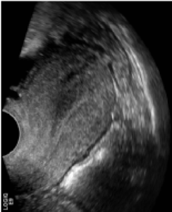
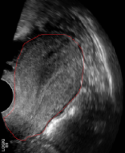
Ultrasound machines use sound waves to generate their images. The sound waves are sent into the body where they bounce off of internal organs and based on how the sound waves return to the machine, a picture is generated. In the above picture, you can see that the uterus is dumbbell shaped with many, different layers. Sometimes an ultrasound gives the doctor all the information that is needed; however, other times a more detailed picture of the uterus is needed to accurately identify the size and location of the fibroids. If the doctor needs more information about the characteristics of the fibroids, an MRI is performed.
MRI
An MRI picture is taken with the patient lying down on a table and passed into the tube-like machine. The MRI machine is very loud, but it is painless and does not use potentially harmful radiation like an x-ray or CT scan does. Depending on the patients’ individual situation, an IV might be inserted into the patient’s arm before the MRI with a special medicine called contrast. Contrast solution may be used to better depict the fibroids. An MRI takes about 30 minutes to perform and the images are crystal clear:
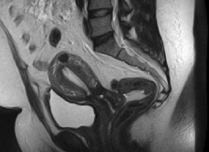
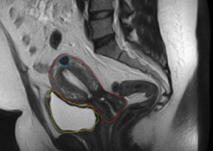
For reference, the right side of the image is the patient’s back, the left side is the front. The uterus is outlined in red on the image on the right. As you can see, the MRI image is much clearer than the ultrasound, giving the doctor a better look, making it easier when diagnosing uterine fibroids. This clearer, more contrasted picture shows the dumbbell shaped uterus, its different layers, and the relationship between the uterus and the surrounding structures (for reference, the bladder is outlined in yellow).
If you look closely at the uterus, you can see a dark object (outlined in blue) towards the top of the uterus; this is a fibroid. This particular fibroid is small and may or may not be causing any symptoms.
In contrast, some patients have many fibroids, as shown in the MRI images below:
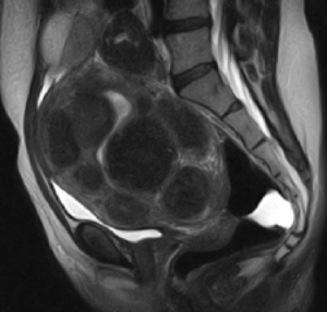
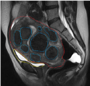
As you can see from the pictures above, the uterus (outlined in red) is larger and more full than the previous pictured uterus. This fullness is a result of multiple fibroids (outlined in blue) within the patients’ uterus. This patient had severe symptoms including excessive menstrual bleeding which caused anemia, pain, constipation, and urinary frequency.
In the MRI picture, you can see how the uterus is squeezing the bladder (outlined in yellow) making it smaller and compressed. Pressure makes the bladder small; therefore, it is unable to entirely fill with urine. When the bladder is unable to properly fill, a woman may experience either the constant urge to pee (even in the middle of the night), or only be able to urinate a small amount at a time due to the squished bladder.
Diagnosing and Treatment Plan
Once the diagnosis of fibroids has been made and the fibroids have been characterized with either an ultrasound and/or an MRI, a treatment plan can be made. A small fibroid like in the first MRI, may not cause enough symptoms to benefit from treatment; however, patients with large or many fibroids typically would.
Uterine Fibroid Embolization
There are many ways to treat fibroids, the gold standard or best option, is called Uterine Fibroid Embolization, or UFE, also known as Uterine Artery Embolization.
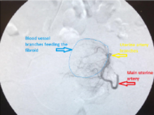
In this procedure, a small plastic catheter is inserted into an artery over the hip or the wrist. Once this plastic catheter is in the artery, IV contrast dye can be injected into the artery. As soon as the dye has had time to distribute, an X-ray picture is taken which opacifies, or “lights up” the artery showing the blood flow.
UFE treatment is an ideal option for women who suffer from fibroids but don’t want to undergo major surgery with a long recovery time. It offers alternatives over a hysterectomy for women who wish to get pregnant in the future. After UFE, your fibroids will shrink, and your toughest symptoms — from heavy menstruation to chronic abdominal pain — can disappear.
In minimally invasive surgery, doctors use a variety of techniques to operate with less damage to the body than with open surgery. In general, minimally invasive surgery is associated with less pain, fewer complications and no hospital stay as it’s done in an outpatient setting.
How UFE works
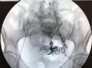
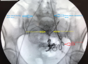
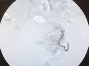
In the first set of pictures below, a catheter is threaded within the squiggly uterine artery and the contrast shows the artery entering the uterus and the fibroids. The doctor can see which artery is feeding the fibroid (the second picture). At this point, small particles can be injected through the plastic catheter and into the uterine artery branches that are supplying the fibroid.
The particles block the arteries which bring blood and oxygen to the fibroid. Without its blood supply, the fibroid will die and shrink down, consequently reducing the symptoms.
Want to learn more about Uterine Fibroid Embolization (UFE) or are in need of a diagnosis?
Give us a call (855) 615-2555 or schedule online.
JACOB WHITE, M.D. – USA FIBROID CENTERS
Dr. Jacob White has been studying and practicing interventional radiology for 10 years. He has conducted research at Drexel Hahnemann University Hospital Radiology, Georgetown University Hospital Radiology, and the National Institutes of Health Radiology. He has contributed extensive research and knowledge to the medical academy through a multitude of presentations and publications. Dr. White is one of 12 doctors on our interventional radiology team. All of our doctors specialize in minimally invasive, non-surgical procedures to treat uterine fibroids.


