


Medically reviewed by
USA Fibroid Centers Medical Review Team
Experienced interventional radiology specialists
Reviewed: July 2026
Pictures of fibroids can paint a visual of what is happening inside your body. Uterine fibroids are non-cancerous growths that develop in or around the uterus, and most women who have them cannot see or feel them without medical imaging. But understanding what fibroids look like, from changes in your body to what imaging reveals, can help you recognize symptoms and know when to talk to a specialist.
The sections below walk through fibroid appearance at every level: what they do to the body from the outside, how they appear on ultrasound and MRI, how different fibroid types and sizes compare, and what imaging shows before and after uterine fibroid embolization (UFE).
What Do Fibroids Look Like?
Fibroids are smooth, firm growths made of muscle and fibrous tissue. They vary widely in appearance depending on their size, location, and how long they have been growing. On imaging, most fibroids appear as well-defined, rounded masses with a different texture than the surrounding uterine muscle. On the outside, fibroids may cause visible changes to the abdomen when they grow large enough to press outward.
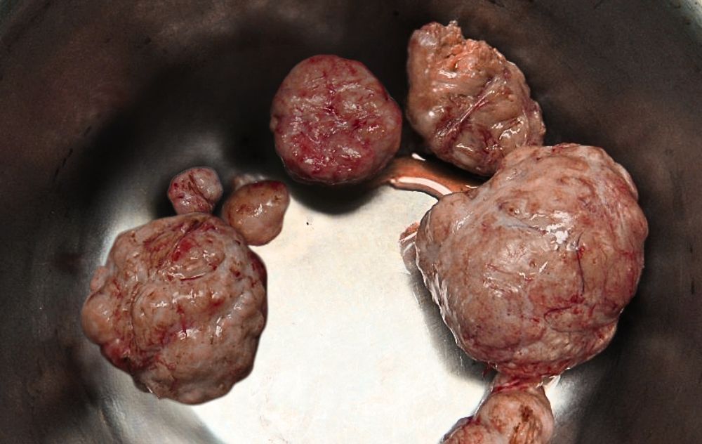
A single fibroid can look very different from multiple fibroids growing together. Some form in clusters that distort the shape of the uterus, while others develop as a single, clearly defined growth attached to the uterine wall. Because fibroids are deeply internal, the clearest pictures of fibroids come from imaging tools, specifically ultrasound and MRI, rather than anything visible from the outside.
What Fibroids Look Like on the Outside: Body Changes

Fibroid Belly and Abdominal Swelling
One of the most common questions women ask is whether fibroids can change how their abdomen looks. The answer is yes, particularly when fibroids grow large or develop in clusters.
A “fibroid belly” refers to abdominal swelling or protrusion caused by one or more large fibroids pressing outward from the uterus. In some cases, the abdomen may appear enlarged in a way that resembles early pregnancy. This occurs most often when fibroids exceed 10 centimeters in diameter or when multiple fibroids are present together.
Women may also notice bloating in the lower abdomen that does not fluctuate with their menstrual cycle the way typical period bloating does. This persistent pressure or fullness can be one of the first visible signs that something is different, and worth discussing with a doctor.
How Fibroids Can Affect Your Appearance Over Time
Fibroids grow slowly in most cases, which means body changes tend to happen gradually. A woman may notice her clothes fitting differently around the waist and hips, or a firm feeling in her lower abdomen when she presses on it.
Larger fibroids may cause the uterus to feel enlarged during a pelvic exam. When a doctor notices an irregularly shaped or enlarged uterus, it is often one of the first steps toward ordering imaging to check for fibroids. For women experiencing changes in abdominal appearance alongside heavy periods, pelvic pain, or pressure, these external signs may point to fibroids that have been growing undetected for some time.
What Fibroids Look Like on Imaging
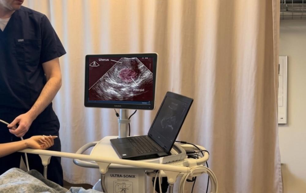
Most fibroids are found through imaging rather than physical examination alone. Two primary tools are used to get pictures of fibroids inside the body: ultrasound and MRI.
How Fibroids Appear on Ultrasound
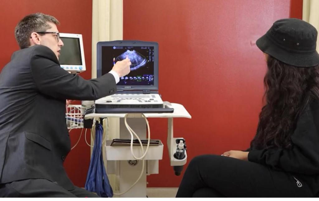
Ultrasound is usually the first imaging test ordered when fibroids are suspected. Depending on the situation, imaging may be performed using an abdominal ultrasound or a transvaginal ultrasound. A transvaginal ultrasound uses a small probe inserted into the vagina to provide detailed, close-up images of the uterus and surrounding structures.
On ultrasound, fibroids typically appear as darker, rounded shapes within or around the uterine muscle. They have a distinct border that separates them from healthy uterine tissue. Ultrasound can show a doctor where fibroids are located, how many are present, and how large they are. However, ultrasound has some limits, as it may miss smaller fibroids or be less accurate in detecting fibroids embedded deep within the uterine wall.
For a thorough assessment, learn more about what goes into a complete fibroid diagnosis evaluation.
How Fibroids Appear on MRI
Magnetic resonance imaging (MRI) may be used in certain situations when additional detail is needed beyond what ultrasound can provide. MRI can help evaluate the size, location, and characteristics of fibroids, particularly when the diagnosis is unclear or when fibroids are difficult to visualize with ultrasound alone. It may also help distinguish fibroids from other conditions, such as adenomyosis, which can appear similar on ultrasound.
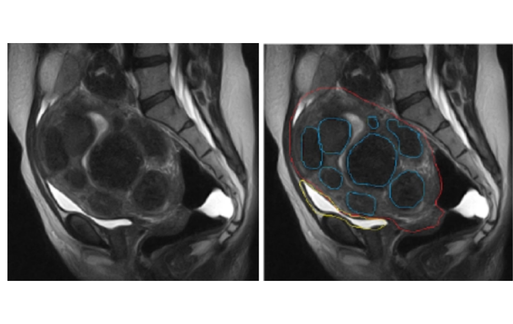
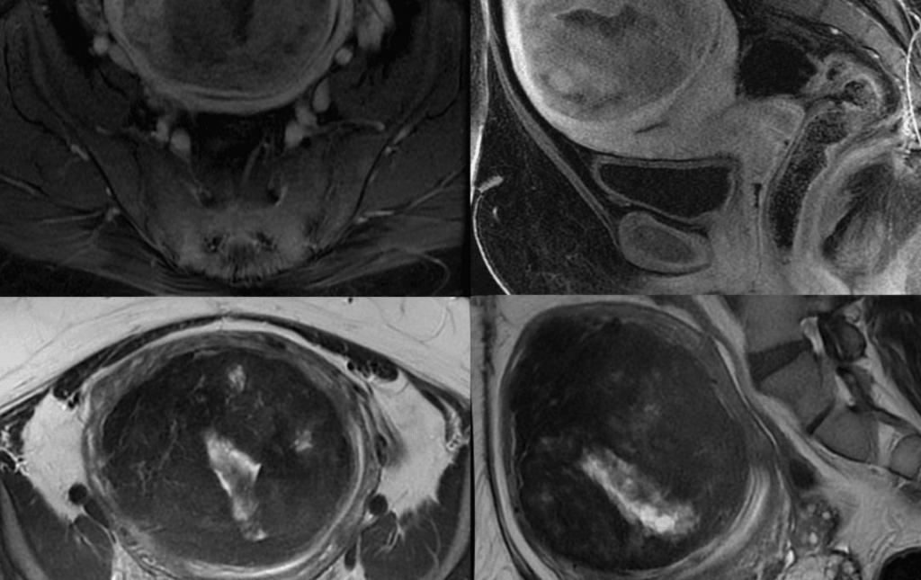
If uterine fibroid embolization (UFE) is being considered, additional imaging may be recommended based on a patient’s individual symptoms, medical history, and treatment goals.
Fibroid Mapping: Getting a Complete Picture
Fibroid mapping is the process of using imaging to document every fibroid’s size, location, and type before treatment. This process helps specialists tailor a treatment plan to each patient’s unique situation.
The imaging used during fibroid mapping may include MRI, ultrasound, or both. Results give a detailed view of the uterus and help identify whether fibroids are inside the uterine cavity, embedded in the wall, or growing outward, all of which influence the symptoms a woman experiences and her options for care.
Image of Types of Fibroids
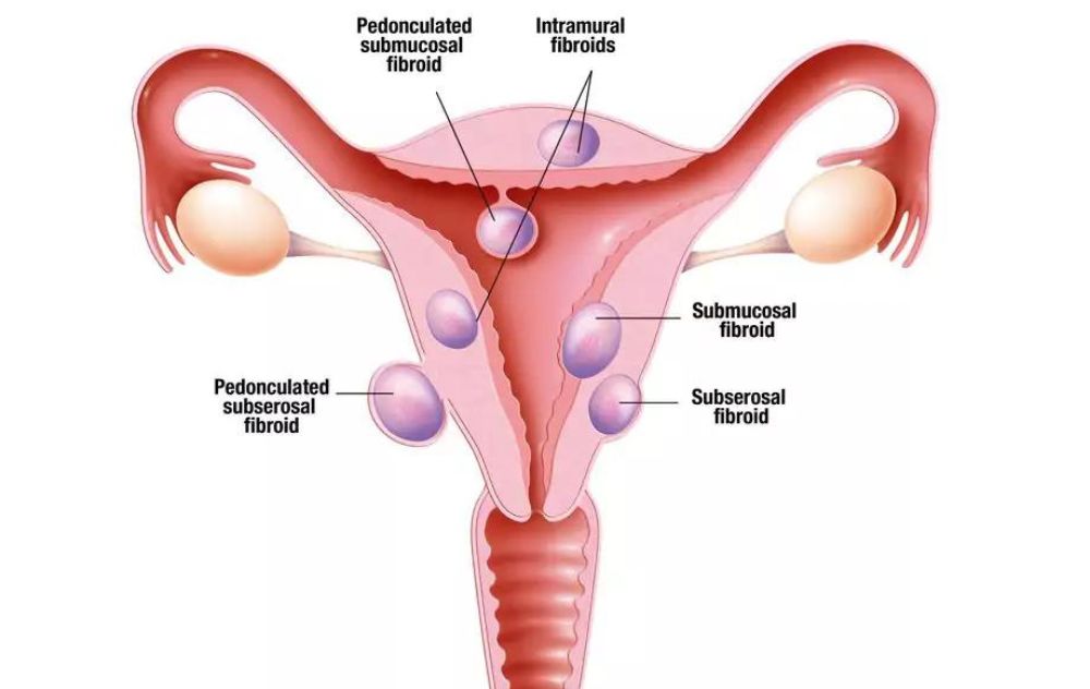
There are several types of uterine fibroids, classified by where they grow in or around the uterus. Each type looks somewhat different on imaging and may cause different symptoms. For a full overview, see our complete guide to types of uterine fibroids.
Intramural Fibroids
Intramural fibroids are the most common type. They develop within the muscular wall of the uterus. On imaging, they appear as rounded, smooth masses embedded in the uterine muscle. When large, they can cause the uterus to appear irregular in shape or larger than normal. Intramural fibroids may cause pelvic pressure, heavy bleeding, and a sensation of fullness in the lower abdomen.
Subserosal Fibroids
Subserosal fibroids grow on the outer surface of the uterus and extend outward into the abdominal cavity. On imaging, they appear as rounded growths attached to the outside of the uterine wall. They can grow quite large before causing symptoms. When very large, they may press against nearby organs, including the bladder or bowel, contributing to frequent urination or bowel discomfort.
Submucosal Fibroids
Submucosal fibroids develop just beneath the inner lining of the uterus, projecting into the uterine cavity. These are associated with some of the most notable symptoms, including very heavy menstrual bleeding and spotting between periods, and can affect fertility. On imaging, submucosal fibroids are visible inside the uterine cavity and are clearly differentiated from the uterine lining.
Pedunculated Fibroids
Pedunculated fibroids are attached to the uterus by a narrow stalk, or pedicle. They can grow into the uterine cavity (submucosal type) or extend outward from the uterine wall (subserosal type). On imaging, the stalk connecting them to the uterus is typically visible and helps distinguish them from other fibroid types. The unique stalk-like structure is a defining characteristic of pedunculated fibroids.
Calcified Fibroids
Calcified fibroids have gone through a process called fibroid degeneration, in which they outgrow their blood supply and begin to break down. Over time, calcium deposits form within the fibroid tissue. On imaging, calcified fibroids often appear brighter on ultrasound and may have a distinctive hardened appearance compared to active fibroids.
Pictures Showing Fibroid Size: From Pea to Grapefruit
Fibroids are often described using fruit comparisons because they vary so dramatically in size. Our detailed fibroid size guide walks through each category in depth.
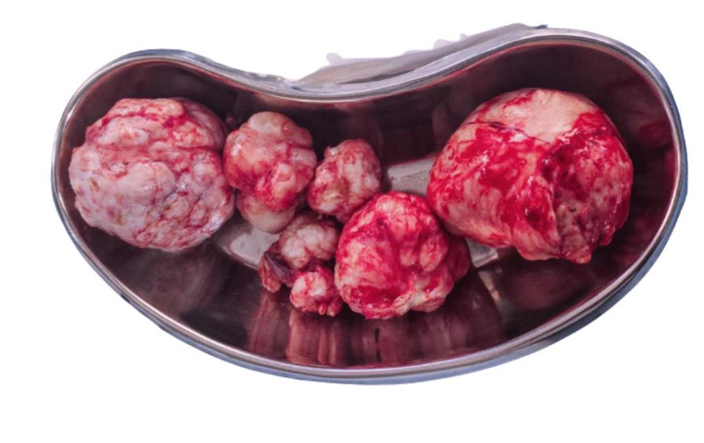
Small fibroids (less than 1 centimeter to 5 centimeters) range roughly from the size of a pea to a cherry. Small fibroids often cause no symptoms and may only be discovered during a routine pelvic exam or imaging done for another reason.
Medium fibroids (5 centimeters to 10 centimeters) are closer in size to a plum or orange. These may begin to cause noticeable symptoms, including heavier periods and pelvic pressure, particularly if they are submucosal or located near sensitive structures.
Large fibroids (greater than 10 centimeters) can be as large as a grapefruit or larger. Fibroids of this size may be more likely to cause significant symptoms or health concerns. They can also press on the bladder or bowel and may cause anemia from heavy fibroid bleeding over time.
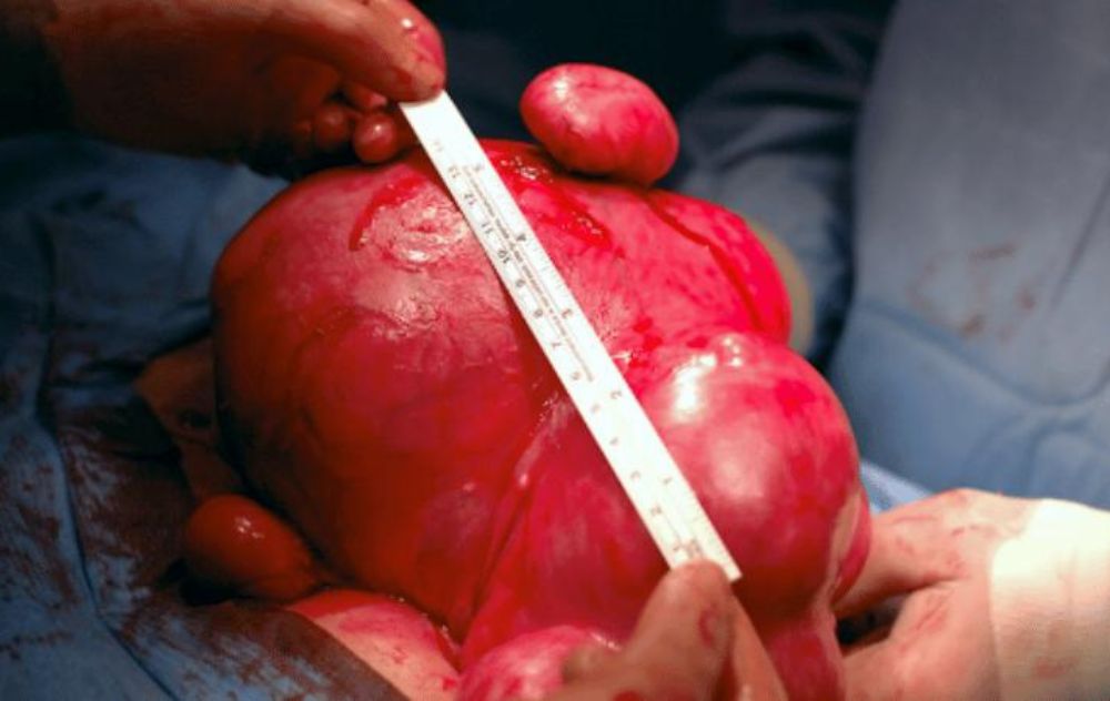
Fibroids can also grow in clusters, with multiple fibroids of varying sizes present at the same time. When this occurs, the combined mass can be much larger than any individual fibroid and may create a more significant impact on surrounding tissue.

If you are looking for diagnostic imaging to determine the size and treatment plan for fibroids, we offer over 40 locations across the USA to help women find clarity and a plan forward.
What Fibroids Look Like Before and After UFE Treatment
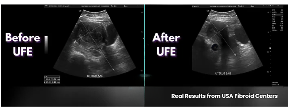
For women who choose uterine fibroid embolization (UFE) as their treatment option, imaging plays an important role not just in diagnosis but in tracking how fibroids respond.
UFE is a minimally invasive, image-guided, outpatient procedure. During the procedure, an interventional radiologist uses imaging in real time to guide a catheter to the arteries supplying blood to the fibroids. Once tiny particles are delivered to cut off that blood supply, fibroids begin to shrink.
Post-treatment MRI and ultrasound images typically show a meaningful reduction in fibroid size over three to six months following the procedure. On imaging, shrinking fibroids may appear smaller, less dense, and less well-defined as they respond to the loss of blood supply. Women who experience a fibroid belly before UFE often notice their abdominal appearance changing as the fibroids reduce in size.
UFE preserves the uterus, is performed without general anesthesia, and allows most patients to return to regular activities within one to two weeks, a meaningful difference from more invasive options.
Behind the Images of Fibroids
Seeing what fibroids look like can help many women visualize the root cause of many of there symptoms. Some women with fibroids do not experience noticeable symptoms and may not realize they have them. However, when symptoms do occur, they are often related to the fibroid’s size, location, and number. Recognizing the signs of fibroids can help understand how they progress, symptoms that you may not have associated with fibroids and mostly important – alert you to when you should seek evaluation and treatment before symptoms begin to affect your daily life.
Common fibroid symptoms include:
- Heavy or prolonged periods: Menstrual bleeding that is heavier than usual, lasts longer than seven days, or requires changing pads or tampons very frequently may be related to fibroids, particularly submucosal types.
- Pelvic pain or pressure: A persistent ache or feeling of fullness in the lower abdomen or pelvis can indicate fibroid growth putting pressure on surrounding tissue.
- Frequent urination: A fibroid near the bladder can press against it, triggering frequent urges to urinate, including during the night.
- Lower back pain: Fibroids pressing on pelvic nerves can cause pain that radiates to the lower back or legs.
- Bloating: Abdominal swelling or a feeling that clothes fit differently, particularly around the waist, may be related to fibroid growth.
- Fatigue and anemia: Heavy fibroid-related bleeding over time can lead to iron deficiency and anemia, causing fatigue, weakness, and difficulty concentrating.
If you are experiencing one or more of these symptoms, it may be worth speaking with a fibroid specialist to determine whether fibroids could be the cause.
Picture Your Future without Fibroids
Seeing what fibroids look like is only part of the picture. Understanding where fibroids are located, how large they are, and whether they may be contributing to symptoms can help you make informed decisions about your care.
If you have questions about your imaging results or would like to learn more about fibroid treatment options, a fibroid specialist can help you better understand your diagnosis and discuss the options that may be right for you.
For a broader visual overview, our visual guide to uterine fibroids covers related ground in depth.
Treatment option: UFE
The Picture of a Fibroid Can Show You the Path to the Right Treatment.
Imaging can reveal what is happening inside the uterus, but understanding what it means for your health takes a specialist who listens. Many women with fibroid symptoms spend years without a clear answer.
UFE is a minimally invasive, outpatient option that treats fibroids without removing the uterus, without general anesthesia, and with a recovery measured in days rather than weeks. A fibroid specialist can review your imaging and help you understand whether UFE may be right for you.
Frequently Asked Questions About What Fibroids Look Like
- Office on Women’s Health, U.S. Department of Health and Human Services, “Uterine Fibroids,” womenshealth.gov/a-z-topics/uterine-fibroids
- National Institute of Environmental Health Sciences (NIEHS), “Uterine Fibroids,” niehs.nih.gov/health/topics/conditions/uterine-fibroids
- Society of Interventional Radiology, “Uterine Fibroid Embolization,” sirweb.org/patients/uterine-fibroids/
Medical Disclaimer: This article provides educational information about uterine fibroids and treatment options. It is not a diagnosis or medical advice. Only a qualified fibroid specialist can determine whether you have fibroids or recommend appropriate treatment. If you have heavy periods, pelvic pain, or other symptoms, consult a healthcare provider or contact USA Fibroid Centers for an evaluation.