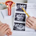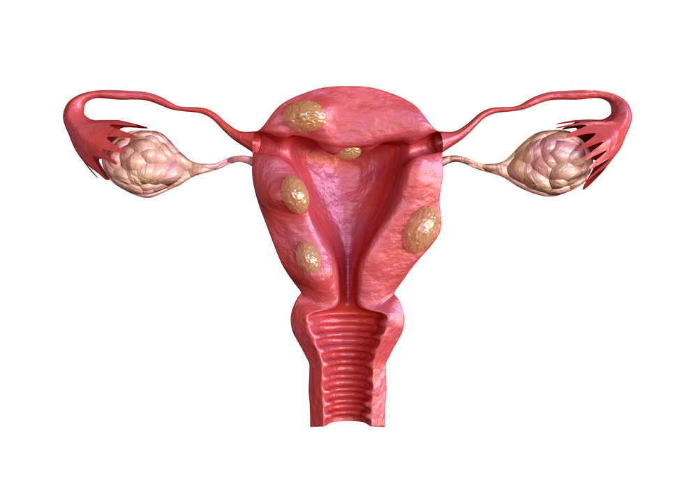
Uterine fibroids can cause a range of symptoms that can significantly impact a woman’s quality of life. While some women may not experience any noticeable signs, others can face debilitating effects. But what do fibroids look like? The following pictures and images of uterine fibroids will help you better understand what fibroids look like, how large they can get, and what symptoms you may experience.
If you’re experiencing symptoms that might be uterine fibroids or other women’s health concerns, such as adenomyosis, consider consulting a doctor specializing in minimally invasive fibroid treatment like the fibroid specialists at USA Fibroid Centers.
Learn More About Treatment Options
The fibroid specialists at USA Fibroid Centers are interventional radiologists who use various imaging tools to diagnose and treat fibroids, such as advanced ultrasound technology, magnetic resonance imaging (MRI), X-ray, or hysteroscopy.
What Do Uterine Fibroids Look Like?
Fibroids are non-cancerous uterine growths nourished by your body’s blood supply. Typically, uterine fibroids appear during a woman’s childbearing years, with African-American women or those with a family history of fibroids being at higher risk.
Fibroids are classified by their location in or on the uterus. Understanding the different locations is important because they impact a woman’s symptoms, treatment options, and potential risks during delivery.
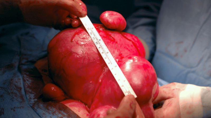
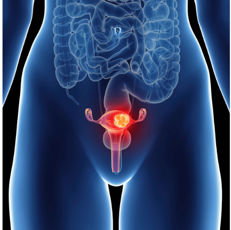
Photo of A Fibroid
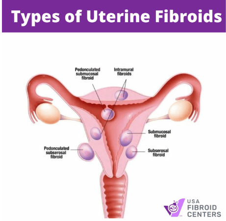
Types of Fibroids
- Intramural: If large, they might grow inside the walls of the uterine muscles and cause pressure, pain, or frequent urination.
- Subserosal: These grow outside the uterus inside the abdominal cavity. They are usually asymptomatic unless very large, which may cause abdominal pain or bloating.
- Submucosal: Projects in the uterine cavity, often causing heavy or irregular periods, and can impact fertility.
- Pedunculated: Extends from the uterine wall by a narrow stalk, allowing it to protrude into the uterine cavity or outside the uterus.
- Calcified: These fibroids outgrow their blood supply and go through fibroid degeneration, causing calcium deposits to develop on the remaining fibroid tissue. Symptoms depend on the fibroid’s location.
By understanding these locations, you can have a more informed conversation with your doctor about your situation and potential treatment options.
SCHEDULE YOUR CONSULTATION TODAY
Fibroid Size Guide
Fibroids can also be classified by their size. These classifications fall under small, medium, and large.
Uterine fibroids vary in size, with some growing to over 10cm, comparable to the size of a mango. Larger fibroids can grow big enough to press against other organs, causing discomfort in the abdomen, back, and pelvic area. They start tiny, around the size of a pea, and increase even reach the size of a small melon.
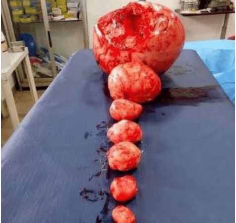
Photo of fibroids ranging in size from small to large.
Small fibroids: Typically less than 1 centimeter in diameter, small fibroids cause minimal symptoms, so treatment might not be necessary immediately but warrants monitoring. Regular checkups with your doctor are recommended to track any potential growth or changes.
Medium fibroids: Around 1 to 5 centimeters in diameter and may cause symptoms such as heavy bleeding, painful periods and pelvic pressure. Depending on the severity of your symptoms, you may be a good candidate for UFE.
Large fibroids: Greater than 5 centimeters in diameter and cause various symptoms, including pelvic pain, pressure on the bladder or bowel, and difficulty getting pregnant. Due to these potential complications, treatment is often recommended.
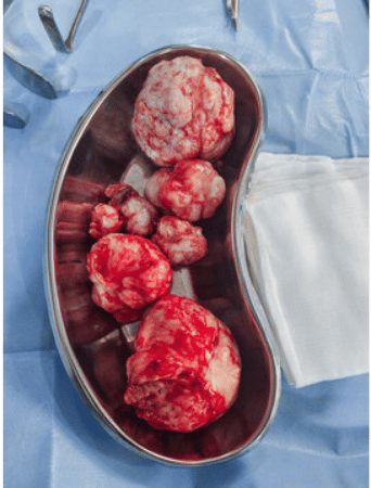
Fibroids grow in various sizes, and the number of fibroids a woman may have differs from individual to individual. The image below shows a uterus with multiple fibroids in and around it.
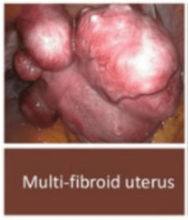
Due to how vastly different one person’s case of uterine fibroids can be from another, consulting a medical professional is necessary for an accurate diagnosis.
Fibroid Diagnosis Imaging
Fibroids are more accurately diagnosed using various tools, such as advanced ultrasound technology, magnetic resonance imaging (MRI), X-ray, or hysteroscopy.
MRI and ultrasound images of fibroids are often used to get a complete picture of their size, location, and type. These uterine fibroid pictures are then used to develop the best treatment for the individual patient, a process known as fibroid mapping.
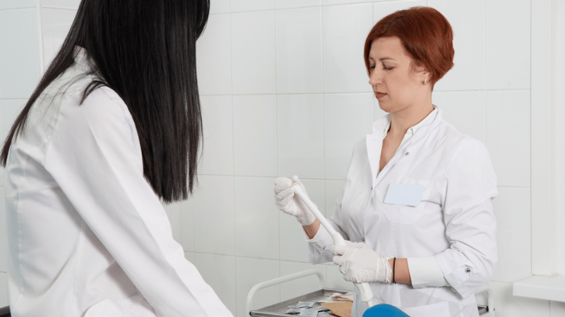
Photo of a transvaginal ultrasound tool, which is used to identify fibroids.
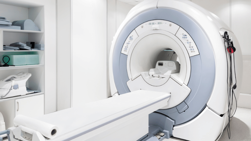
Photo Caption: An MRI Machine
Bird’s Eye View: How Imaging Shows Fibroid Size and Location
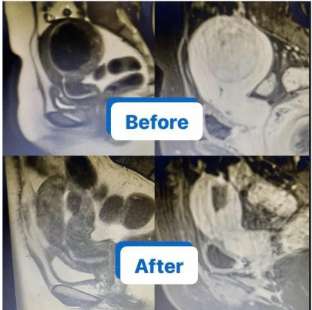
The image above is an MRI showing the uterus before and after UFE treatment.
Looking closely at the uterus in the image below, you can see a dark object (outlined in blue) towards the top. This dark object is a fibroid. The patient had severe symptoms, including excessive menstrual bleeding, which caused anemia, pain, constipation, and urinary frequency.
Below are more images fibroid images from a diagnosis consultations using ultrasounds, MRIs, and X-rays.
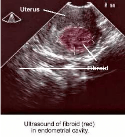
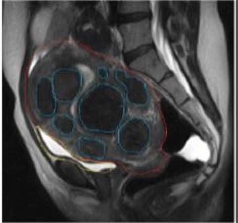
These pictures of uterine fibroids showcase their impact on surrounding tissues and organs. It is important to understand their location in and around the uterus and associated symptoms.
If you are experiencing symptoms such as pelvic pain, bloating, or heavy menstrual bleeding, you may have fibroids contact USA Fibroid Centers to schedule a screening, discuss your concerns, and explore treatment options, including minimally invasive treatment with uterine fibroid embolization (UFE).
Get an Accurate Diagnosis: Contact Us Today
Fibroids 101
Let’s begin with the basics of understanding a woman’s reproductive system and how fibroids can impact it.
The uterus is a pear-shaped organ in the lower abdomen, between the bladder and the rectum. It is the area that carries a developing fetus during pregnancy and composes three layers of muscle: the endometrium, the myometrium, and the perimetrium.
Photo caption: Illustration depicting the uterus
The uterus looks like a light bulb with wings curling back into the uterus. The uterus diagram above shows these wings comprise the Fallopian tubes and ovaries. Meanwhile, the uterus and cervix (leading into the vagina) are on the top and bottom of this “lightbulb” of your body.
Uterine Fibroids: Don’t Ignore the Symptoms
Uterine fibroids are uterine growths that can cause a range of symptoms. While the severity depends on the location, size, number, and type of fibroids, some common signs to watch out for include:
- Heavy or Lengthy Periods: This is one of the most frequent symptoms, with periods lasting longer than usual and bleeding being heavier than normal.
- Bloating and Abdominal Discomfort: You might experience swelling in your lower abdomen or belly, similar to pregnancy bloating.
- Pelvic Pain and Pressure: A persistent ache or pressure in the pelvic area can be a sign of fibroids.
- Lower Back and Leg Pain: Fibroids can sometimes press on nerves, causing pain in your lower back or radiating down your leg.
- Frequent Urination: A fibroid near your bladder can cause a frequent urge to urinate, even at night.
- Constipation: Large fibroids might press on your bowels, leading to constipation issues.
- Painful Intercourse: Fibroids can sometimes affect sexual intercourse, causing discomfort or pain.
- Anemia: Heavy bleeding over time can lead to iron deficiency and anemia, causing fatigue and weakness.
If you’re still unsure whether you have fibroids, our symptom checker can help you determine whether you should consult a fibroid specialist for treatment.
Fibroid Treatment
Uterine Fibroid Embolization (UFE) is a minimally invasive, short outpatient procedure for treating fibroids. A tiny catheter is inserted into the uterine artery, feeding the fibroid and injecting embolic agents. The catheter then blocks blood flow, causing the fibroids to shrink and die.
UFE preserves the uterus, allowing women to maintain their fertility. This treatment also has a lower risk of complications and a shorter recovery time than surgery. Additionally, UFE effectively treats multiple fibroids simultaneously, regardless of their size or location, providing long-term symptom relief.
When to Seek Uterine Fibroid Treatment
Although many women have fibroids, not everyone realizes they have them. Symptoms can range from mild to severe and are typically associated with a bad period.
If you are experiencing any combination of the common fibroid symptoms, it’s time to schedule an appointment with your doctor to figure out if you have fibroids and what the best path forward is for treatment.
If you’re concerned that you may have fibroids or want an alternative to invasive surgery, contact USA Fibroid Centers to request a consultation online or call us at 855-615-2555. We accept most insurances and offer self-pay options at our locations across the United States.
No matter where we meet, we look forward to helping you regain control of your life.

